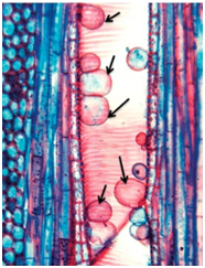
The balloon-shaped structures called tyloses
1. are linked to the ascent of sap through xylem vessels
2. originate in the lumen of vessels
3. characterize the sapwood
4. are extensions of xylem parenchyma cells into vessels
In the given diagram of the root apical meristem, what would be true for the structure labeled as number 1?
1. The cells in the region continually divide to produce cells of region 5
2. The cells in this region divide at much faster rate than the cells of region 2
3. The cells of this region are able to repopulate the borderline meristematic regions when they are injured
4. The cells of this region include the initials of region 3
The given diagram shows the central core of root in a certain plant. P is phloem and protoxylem elements and V is metaxylem. What is correct for structures indicated by arrows?
| 1. | They show aerenchyma essential for aeration of root cells |
| 2. | They show cells in the roots that contain chloroplast |
| 3. | They show cells lack the characteristic radial and inner tangential wall thickening which is common to endodermal cells |
| 4. | They show cells that store resins and oils in the roots |
The balloon like structures seen in vessels in the given diagram are called as:

1. Tyloses
2. Hydathodes
3. P proteins
4. Lenticells
What type of vessels are you most likely to see in metaxylem of a stem?
| 1. |  |
2. |  |
| 3. |  |
4. |  |
Identify the correct statement regarding A and B [types of concentric vascular bundles] in the given diagram:
| 1. | A is Amphivasal and found in Dracaena while B is amphicribal and found in Selaginella |
| 2. | A is Amphivasal and found in Selaginella while B is amphicribal and found in Dracaena |
| 3. | A is Amphicribal and found in Dracaena while B is amphivasal and found in Selaginella |
| 4. | A is Amphicribal and found in Selaginella while B is amphivasal and found in Dracaena |
Vascular bundles are continuous from the root to the stem, but different arrangements are found in two cases. What would be true for this arrangement?
| 1. | In the stem they are collateral with endarch xylem while in the root they are radial with exarch xylem |
| 2. | In the stem they are collateral with exarch xylem while in the root they are radial with endarch xylem |
| 3. | In the stem they are radial with endarch xylem while in the root they are collateral with exarch xylem |
| 4. | In the stem they are radial with exarch xylem while in the root they are collateral with endarch xylem |
If you look at the section of a three year old eudicot stem, the vascular bundles will show:
| 1. | 3 rings of xylem and 3 of phloem. |
| 2. | 2 rings of xylem and 1 of phloem. |
| 3. | 2 rings of xylem and 3 of phloem. |
| 4. | 3 rings of xylem and 1 of phloem. |
In the section of a monocot leaf shown in the given diagram, what would be true for the structures labeled X?
| 1. | They regulate the opening and closing of the stomata in the adaxial surface of the leaves |
| 2. | Due to presence of lignin in their walls they provide support to the leaf surface in windy conditions |
| 3. | They are involved in folding and unfolding of leaf tissue to control light intensity and reduce overall water loss |
| 4. | They are abnormal cells produced during healing of the adaxial surface after injury and are common portals of entry of viruses into the plants |
The diagram shows the development of lateral roots. This development will be called as:
1. Exogenous
2. Endogenous
3. Exarch
4. Endarch


